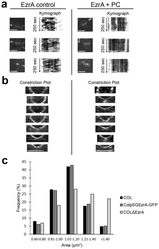
Extended Data Figure 7. Z-ring protein EzrA shows impaired treadmilling in the presence of PC190723 and biphasic ring constriction.
a, ColpSGEzrA-GFP cells, expressing a functional EzrA fusion to GFP, were imaged by SIM every 5 sec in the absence (EzrA control) or presence (EzrA + PC) of PC190723. Kymographs were obtained by extracting fluorescence intensity values along the black line indicated in cells in the left panels. Similarly to what was observed for FtsZ55-56sGFP, addition of PC190723 abolished EzrA movement (vertical lines in the kymographs). b, ColpSGEzrA-GFP cells were imaged by SIM every 5 min in the absence (left) or presence (right) of PC190723 and kymographs showing constriction of EzrA-GFP rings were plotted. In control conditions the larger EzrA rings showed biphasic constriction behaviour while in the presence of PC190723 only the rings in the second stage of cytokinesis were able to constrict. Data in (a, b) are representative of two biological replicates. Scale bars, 1 μm. c, To test the functionality of the EzrA-GFP construct, strains COL, ColpSGEzrA-GFP and COLΔEzrA (lacking ezrA) were imaged by phase contrast and cell area was measured. The lack of EzrA in COLΔEzrA (N=959) resulted in cell enlargement, while the size distribution of ColpSGEzrA-GFP (N=957) cells mimicked that of parental strain COL (N=851), indicating that the EzrA fluorescent fusion is functional.

