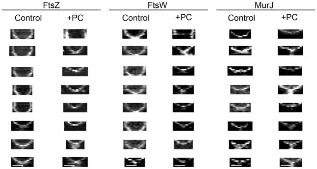
Extended Data Figure 8. Kymographs showing constriction of FtsZ55-56sGFP, FtsW-sGFP and MurJ-sGFP rings during cell division.
Strains ColFtsZ55-56sfGFP, ColFtsW-sGFP and ColMurJ-sGFP were imaged every 10 min in the absence (control) or presence (+PC) of PC190723 for a total of 60 min. MurJ-sGFP control kymographs were performed on cells where MurJ-sGFP signal appears on the second frame, to ensure that the entire constriction process was observed (i.e. that the absence of a biphasic behaviour did not result from only imaging cells in later stages of cell division). FtsZ55-56sGFP and FtsW-sGFP rings showed a biphasic constriction behaviour (no/slow constriction followed by fast constriction). Addition of PC190723 inhibited constriction of larger FtsZ55-56sfGFP and FtsW-sGFP rings (see top kymographs), but not of smaller rings which were undergoing fast constriction. MurJ-sGFP rings only displayed fast constriction and therefore were always able to constrict in the presence of PC190723. Data are representative of three biological replicates. Scale bars, 0.5 μm.

