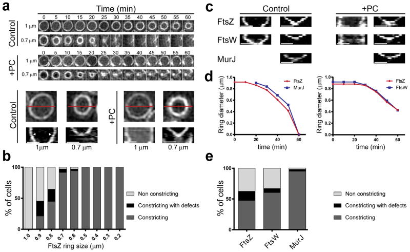
Figure 5. PG synthesis provides force for septum constriction.
a, Timelapse images and corresponding kymographs of FtsZ55-56sGFP rings in the absence (control) or presence (+PC) of FtsZ inhibitor PC190723. Kymographs were obtained by drawing a line (exemplified in red) across the cell for each time point. Arrowheads point to transition from first (no/slow constriction) to second (fast constriction) step of cytokinesis. b, Percentage of FtsZ55-56sGFP rings of different diameters that constricted in the presence of PC190723 (N=248). c, Kymographs of FtsZ55-56sGFP, FtsW-sGFP and MurJ-sGFP larger (left) and smaller (right) rings. PC190723 only abolished constriction of larger rings. d, Graphs showing diameter of FtsZ-mCherry and MurJ-sGFP rings (left) or FtsZ-mCherry and FtsW-sGFP rings (right) in single cells during cytokinesis. e, Percentage of FtsZ55-56sGFP, FtsW-sGFP and MurJ-sGFP rings that constricted in the presence of PC190723 (FtsZ N=254; FtsW N=176; MurJ=188). Data in (a, c) are representative of three biological replicates. Scale bars, 0.5 μm.

