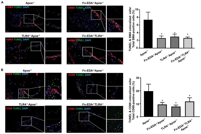
Figure 3.
FN-EDA promotes VSMCs and macrophage apoptosis in advanced innominate lesions via TLR4. Left-side show representative immunofluorescence images of sections stained with A, TUNEL, and αSMA. B, TUNEL and CD68. Nuclei were visualized with DAPI stain. Boxed region in A & B is magnified to show colocalization of TUNEL/αSMA-positive cells (A) and TUNEL/CD68-positive cells (B). Right side shows quantification of TUNEL/αSMA-positive cells and TUNEL/CD68-positive cells. Data represent mean ± SEM. *P<0.05 versus Apoe−/− mice. N=7–8 mice/group. Value for each mouse represents a mean from 4 serial sections (each section approximately 60 μm apart). Statistical analysis: Figure 3A; Kruskal-Wallis test followed by uncorrected Dunn’s test. Figure 3B; Parametric one-way ANOVA followed by Sidak’s multiple comparisons test. Scale bar= 50 μm.

