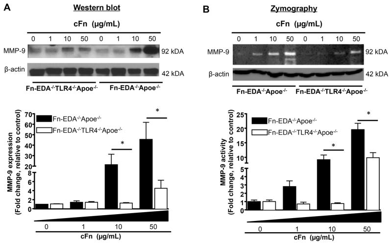
Figure 6.
Dose-dependent effect of exogenous cFn on MMP9 activity in macrophages. Pooled bone marrow-derived macrophages from Fn-EDA−/−Apoe−/− and Fn-EDA−/−TLR4−/−Apoe−/− mice (n=5 mice/group) were stimulated in the presence of cFN (0–50 μg/mL) for 24 hours. A, Top panel shows representative Western blots showing MMP9 expression and β-actin (loading control). Bottom panel represents quantification of the intensity of MMP9 to β-actin. N = 5 experiments/group. B, Top panel shows MMP9 activity (gelatin zymography). The same amount of protein was loaded on another gel and stained for β-actin (loading control). The bottom panel shows quantification of the intensity of MMP9 normalized to β-actin in panel A. Data is presented as mean ± SD. *P<0.05. N = 5 experiments/group. Statistical analysis: Two way ANOVA followed by Holm-Sidak multiple comparison tests.

