Figure 11.
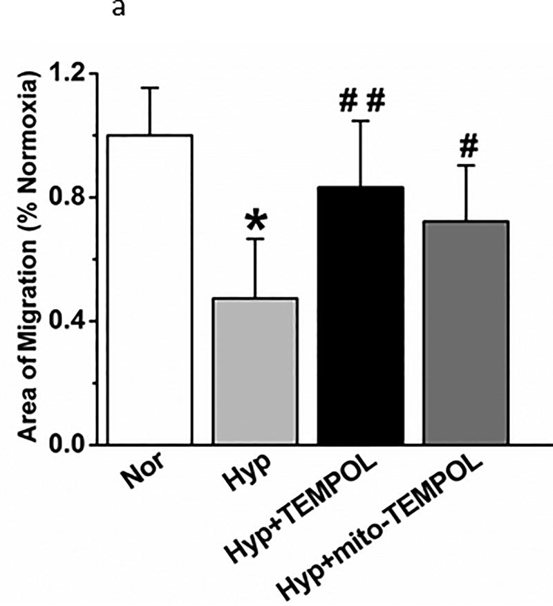
(a). Hyperoxia decreased migration of cells into a path cleared by a scratch relative to counterparts maintained in normoxia. TEMPOL or mito-TEMPOL partially restored diminished migration of PECs in hyperoxia by 48 hours. Data were not normally distributed and were compared by ANOVA on Ranks. *P < 0.05, different from normoxia, #P < 0.05 different from hyperoxia; ##P < 0.01, different from hyperoxia. n=12 experiments for each condition and time.
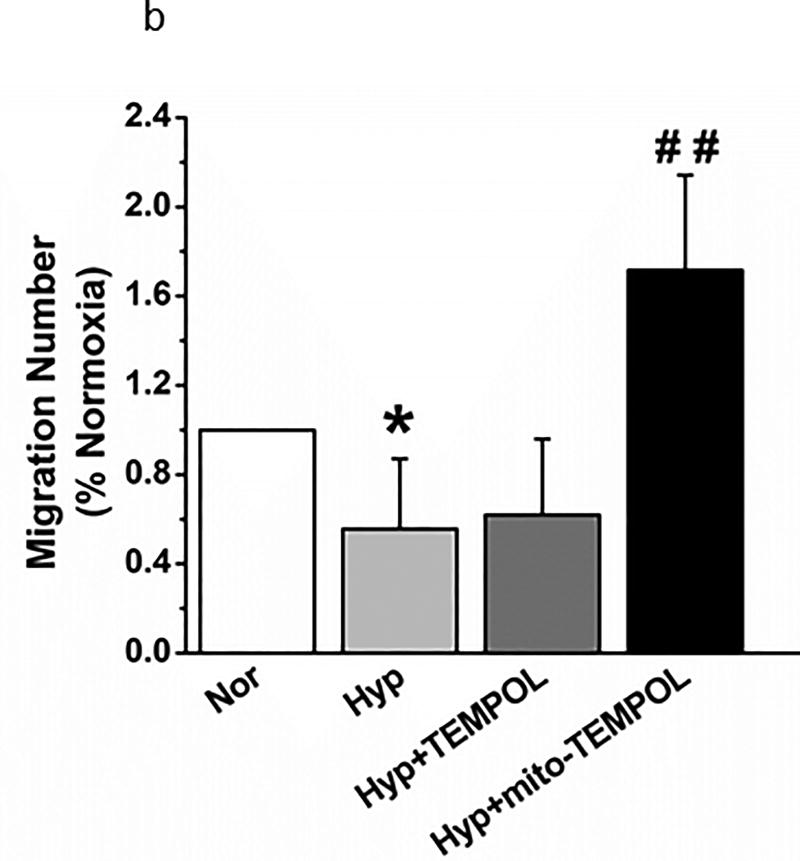
(b). Transwell migration of PECs was compared in normoxia and hyperoxic conditions with 4 separate isolates of endothelial cells. 48 hours hyperoxia decreased migration. Mito-TEMPOL but not TEMPOL restored migration to supra-normoxia values. (n=29,29,23, and 22 wells respectively for each of the above 4 groups from 4 separate experiments. Data were normally distributed with equal variance and compared by ANOVA followed by Dunnett’s. *P < 0.05, different from normoxia, ##P < 0.01, different from hyperoxia.
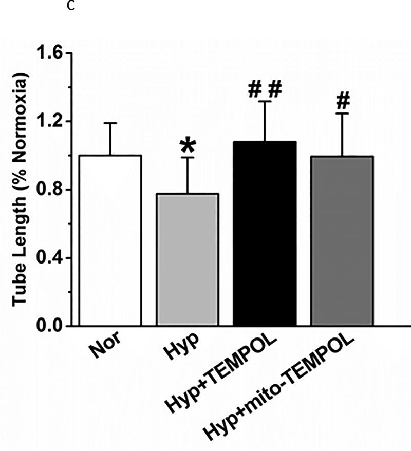
(c). Network formation of PECs in a 3-dimensional matrix is decreased by 24 hours hyperoxia. Both TEMPOL and mito-TEMPOL restore network formation. (n=17–21 fields/condition, from 4 separate experiments). Data were normally distributed with equal variance and compared by ANOVA followed by Dunnett’s. *P < 0.05, different from normoxia; #P < 0.05 and ##P < 0.01 different from hyperoxia.


