. 2018 Feb 5;59(2):341–344. doi: 10.3349/ymj.2018.59.2.341

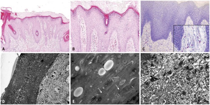
Fig. 2. Skin biopsy features. (A and B) Hyper- and parakeratosis, hypergranulosis, irregular acanthosis and follicular plugging. Dilated vessels with inflammatory cellular infiltrates of lymphocytes, histiocytes and eosinophils in the upper dermis (hematoxylin-eosin, ×100 and ×200, respectively). (C) Increased number of mast cells in the upper dermis (toluidine blue, ×400). (D) Electron microscopy shows numerous lipid droplets in the corneocytes. Parakeratotic corneocytes are also noted (×2K). (E) Electron-lucent lipid droplets are observed, but corneodsmosomes are well preserved (arrows) (×40K). (F) Increased number of polysomes in the keratinocytes of the upper spinous layer (×20K).

Fig. 2