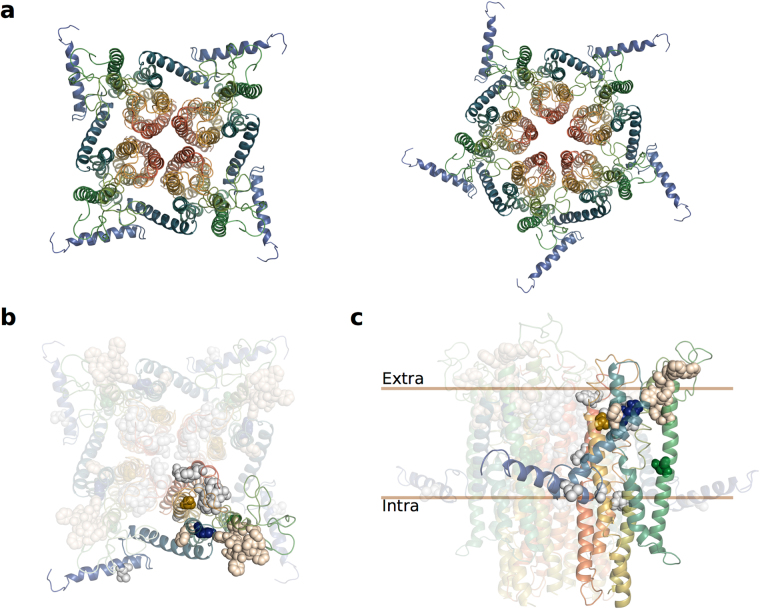
Figure 6.
(a) 3D models of putative Mayetiola destructor (Mdes) Orco tetra- and pentamers, viewed from the extracellular surface. (b) Putative tetramer model of MdesOrco multimer, viewed from extracellular surface, with selected residues shown in sphere representation. Blue (H81F), green (N133L) and gold (S350A) spheres represent MdesOrco residues mutated in this study. Grey spheres represent corresponding residues associated with cation selectivity or ion channel function; tan spheres represent residues associated with ligand binding34. (c) Side view of MdesOrco tetramer with selected residues shown in sphere representation as described in (b). Note: protein models are coloured from blue (N-termini) to red (C-termini).

