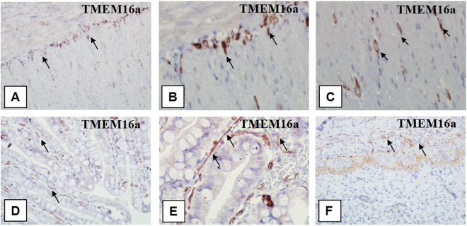
Figure 6.
Immunohistochemical staining of the bowel wall with primary antibodies to TMEM 16a (positive control). (А–Е) The bowel wall (of a five-day-old newborn). Strong telocyte staining is detected in the smooth layer. Telocytes form enclosed structures and capillary walls in the same places. (A) х200. (B) x400. (C) x600. (D) and (Е) The presence of telocytes in the crypts and different layers (indicated with arrows). (D) х200. (E) x400. (F) The small intestine wall (of a foetus at the 20th week of gestation). Weak immunohistochemical staining of telocytes is detected in the muscle layer, x200.

