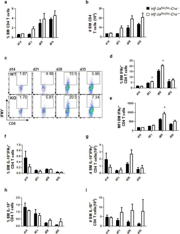
Figure 3.
HIF-1α deficiency in CD11c+ cells results in stronger Th1 responses in the bone marrow. Hif-1αflox/floxCd11c-Cre− (WT) and Hif-1αflox/floxCd11c-Cre+ (KO) mice were infected with 2 × 10⁷ LV9 amastigotes intravenously. (a) Graph represents the frequency and (b) absolute numbers of bone marrow CD4 T cells. (c) Representative FACS plots for IFNγ+CD4 T cells of Hif-1αflox/floxCd11c-Cre− (upper panels) and Hif-1αflox/floxCd11c-Cre+ mice (lower panels) in the bone marrow. (d) Percentage and (e) mean fluorescence intensity (MFI) of IFNγ+CD4 T cells in the bone marrow. Frequency (f) and numbers (g) of IFNγ+IL-10+ double producing CD4 T cells; percentage (h) and numbers (i) of IL-10+ single producers CD4 T cells in the bone marrow over the course of infection. All data represent mean ± SEM of one of 3 independent experiments, n = 4.

