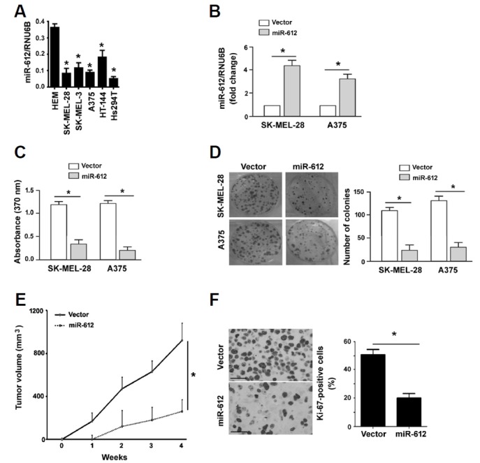
Fig. 2. Restoration of miR-612 reduces proliferation and tumorigenesis.

(A) Measurement of miR-612 levels in human epidermal melanocytes (HEM) and melanoma cell lines. *P < 0.05 relative to HEM. (B) qRT-PCR analysis of miR-612 expression in melanoma cells transfected with vector or miR-612-expressing plasmid. *P < 0.05. (C) BrdU incorporation assays were performed to assess the proliferation of melanoma cells transfected with vector or miR-612-expressing plasmids. (D) Colony formation assay. Melanoma cells stably transfected with vector or miR-612-expressing plasmids were cultured for 10 days to allow to form colonies. Representative images of colonies are shown in left panels. (E) Xenograft tumor studies. Subcutaneous tumors derived from miR-612-overexpressing A375 melanoma cells grew significantly slower than control tumors (n = 4). (F) Ki-67 immunohistochemistry. Sections of xenograft tumors were stained with anti-Ki-67 antibody. Right, quantification of Ki-67 immunostaining. Scale bar = 50 μm. *P < 0.05.
