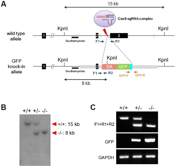
Fig. 4. Generation of Redrum-targeted mouse model.

Map of the wild type Redrum locus is shown at the top, and the locus after homologous recombination is shown below (A). A Kpn I site is introduced by homologous recombination and would result in 8 kb fragment upon Kpn1 digestion. Locations of the probe for Southern blotting and primers (F1, R1, R2, GFP-F, and GFP-R) for PCR amplification are indicated (A). Southern blotting with genomic DNA derived from wild type, heterozygous and homozygous mice shows results consistent with the scheme of the homologous recombination (B). Likewise, PCR amplification resulted in the predicted pattern (C). (SA, splice acceptor; GFP, green fluorescent protein; GAPDH, Glyceraldehyde 3-phosphate dehydrogenase)
