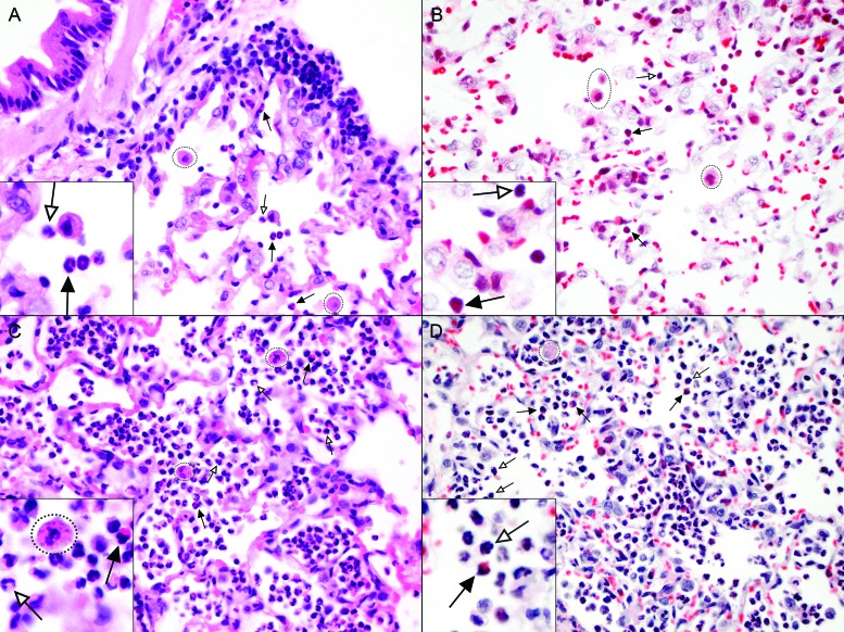
Figure 2.
Standard staining with hematoxylin and eosin is better than Luna staining for visualization of cotton rat eosinophils in lung tissue. Paraffin-embedded lung sections from cotton rats (A and B) 4 d after infection with RSV or (C and D) 1 d after infection with S. aureus were stained with (A and C) hematoxylin and eosin or (B and D) Luna stain. Variable numbers of eosinophils with bilobed nuclei and prominent pink to red cytoplasmic granules (black arrows), neutrophils with segmented nuclei and less distinct cytoplasmic granules (white arrows), and alveolar macrophages (dotted circles), are present within alveoli. Macrophages in S. aureus infected cotton rats frequently contain cocci. Magnification, 60×; insets, 100×.

