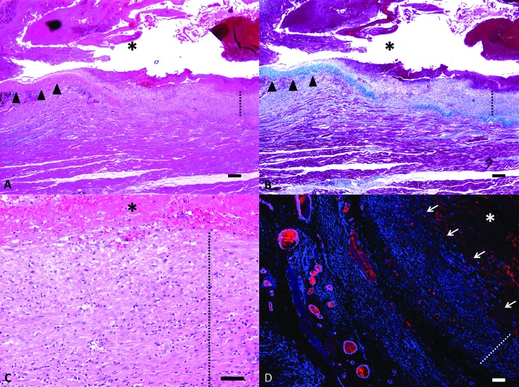
Figure 6.
Microscopic findings. (A) The pseudoaneurysm is composed of a large blood-filled cavity (asterisk) that is surrounded by a capsule composed segmentally of dense collagen (arrowheads) and smooth muscle (dotted line). Hematoxylin and eosin stain; bar, 200 μm. (B) Masson trichrome staining highlights the capsular segments composed of collagen (blue, arrow heads) and smooth muscle (dotted line). (C) Magnified image illustrating the layering of the smooth muscle cells (dotted line), similar to normal tunica media, in segments of the capsule. Hematoxylin and eosin stain; bar, 50 μm. (D) Immunofluorescent staining for α-smooth muscle actin and von Willebrand factor highlight the smooth muscle within the wall of the capsule (dotted line) and the lack of an endothelial lining (arrows). Confocal microscopy; bar, 100 μm.

