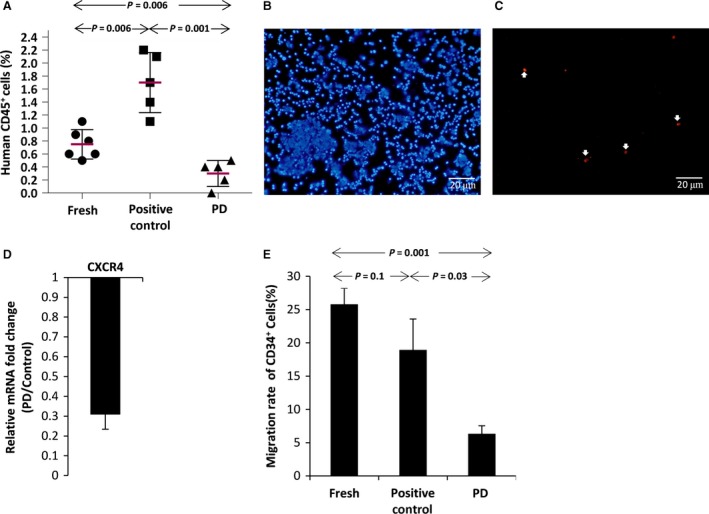
Figure 5.

(A) Percentage of human CD45 positive cells in peripheral blood of newborn mice after one‐week treatment with human haematopoietic growth factors [SCF (4 ng/g), IL3 (4 ng/g) and G‐CSF (50 ng/g)]. (B) Immunocytochemistry to identify human cells in the bone marrow of newborn mice, DAPI stains nucleus (Blue), (C) human nuclear antibody (Red). (D) CXCR4 gene expression in PD‐expanded CD34+ cells relative to the positive control group detected by quantitative real‐time PCR (n = 3). (E) Percentage of CD34+ expanded cells migrated through the transwell in response to SDF‐1 as a chemoattractant factor.
