
Figure 6.
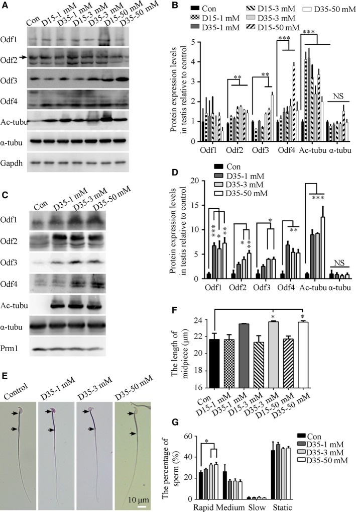
Effect of lithium on the formation of ODFs, midpiece and sperm speed in vivo. (A) Representative Western blots for the expression of Odf family proteins, acetylated α‐tubulin and total α‐tubulin in testes treated with LiCl at different doses and time courses. D indicates days. (B) Quantification of band intensity for Odf family proteins, acetylated α‐tubulin and total α‐tubulin in panel A by Western blot analysis. The expression of Odf2, 3, 4 and the acetylation of α‐tubulin were increased significantly by LiCl (n = 3 experiments). (C) Representative Western blots for the expression of Odf family proteins, acetylated α‐tubulin and total α‐tubulin in sperm treated with LiCl at different doses and time courses. D indicates days. (D) Quantification of band intensity for Odf family proteins, acetylated α‐tubulin and total α‐tubulin in panel C by Western blot analysis. The expression of Odf1, 2, 3, 4 and the acetylation of α‐tubulin were increased significantly by LiCl (n = 3 experiments). (E) Representative images of sperm after LiCl treatment with the midpiece length labelled. Black arrows indicate the start and end points of midpiece. (F) Quantification of the midpiece length of sperm after LiCl treatment at different doses and time courses (N = 3 males per group; n ≥ 200 sperm per male). The midpiece length in sperm from 3 groups that were treated with 3 and 50 mM LiCl for 35 days was significantly longer than those in the control group. Note the truncated y‐axes. Scale bar = 10 μm. (G) Statistical results were shown the distribution of motile types of sperm after LiCl treatment at different doses and courses (N = 3 males per group). The data are presented as the means ± S.E.M. Student's t‐test was used for the statistical analysis. Statistical significance is expressed relative to the untreated control. *P < 0.05; **P < 0.01; ***P < 0.001; NS, P > 0.05.