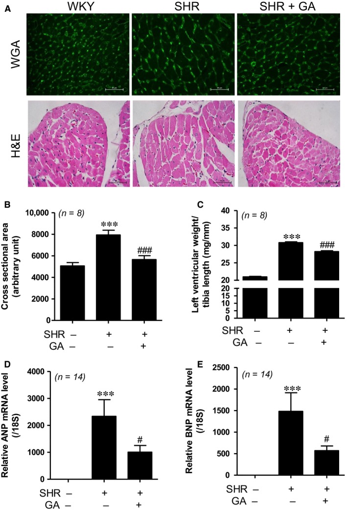
Figure 2.

Gallic acid attenuates left ventricular hypertrophy in spontaneously hypertensive rats (A) WGA (top panel) and H&E (bottom panel) staining performed to evaluate the increased size in myocytes. Scale bar = 50 μm. (B) Cross‐sectional area of the left ventricle was evaluated (n = 8 per group). ***P < 0.001 compared with WKY rats. ### P < 0.001 versus SHRs. (C) The ratio of the left ventricular weight to tibia length (mg/mm) in WKY, SHR, SHR+GA (n = 8 per group). ***P < 0.001 compared with WKY rats. ### P < 0.001 versus SHRs. (D, E) The mRNA levels of ANP and BNP were evaluated by real‐time RT‐PCR from three groups (n = 14). The transcript levels were normalized to those for 18S and presented as relative values. ***P < 0.001 versus WKY rats. # P < 0.05 versus SHRs.
