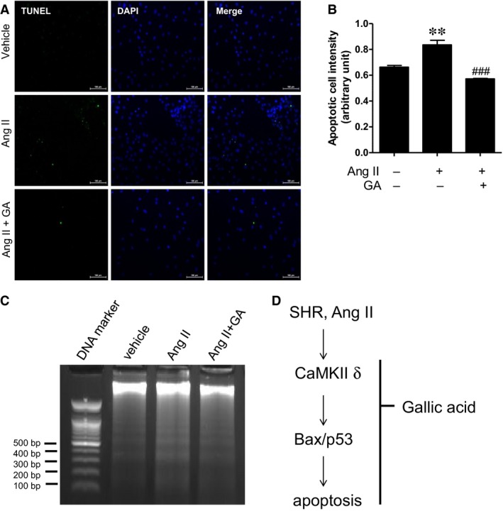
Figure 6.

Gallic acid reduces angiotensin II‐induced apoptosis as determined by TUNEL assay and DNA fragmentation. (A) Representative images of TUNEL staining. H9c2 cells were serum starved for 24 hrs and were treated with gallic acid (50 μM) in the presence or absence of angiotensin II (10 μM) for 36 hrs. Green and blue colours indicate apoptotic cells and nuclei in H9c2 cells. (B) Quantification of positive TUNEL staining. **P < 0.01 versus vehicle group. ### P < 0.001 versus the angiotensin II‐treated group. Data represent the means ± S.E. of at least three independent experiments. (C) Representative DNA fragmentation of angiotensin II (100 μM)‐treated H9c2 cells in the presence or absence of gallic acid (25 μM). H9c2 cells lysates were treated with RNase A and proteinase K before DNA extraction. Apoptosis was determined by 1.7% agarose gel electrophoresis; 100 bp DNA ladder was used as a loading control.
