
Figure 6.
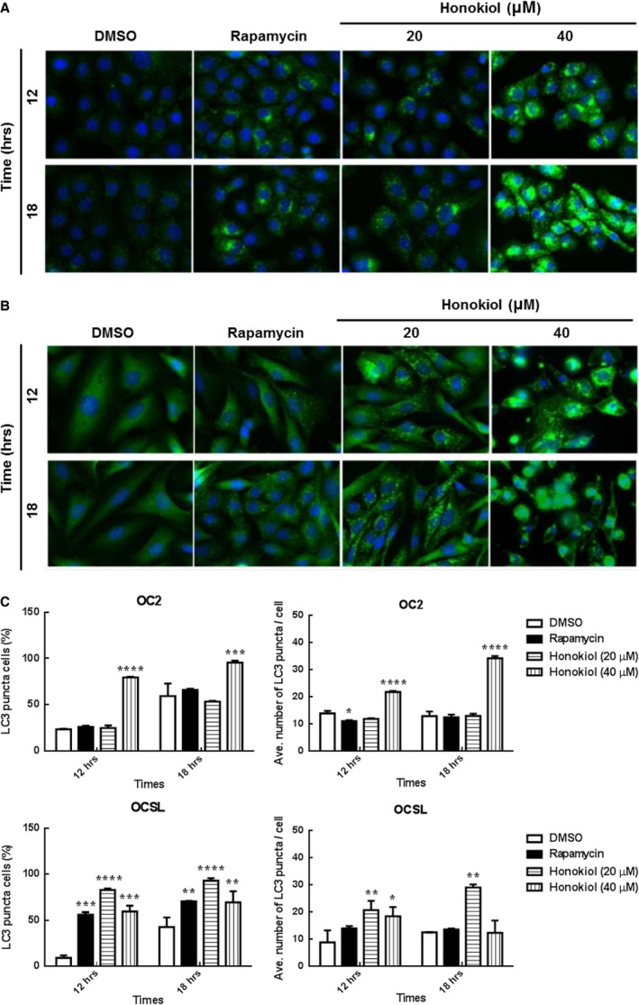
Autophagy was triggered after honokiol treatment in human OSCC cells. (A) OC2 and (B) OCSL cells were treated with DMSO, rapamycin and honokiol for 12 and 18 hr. Cells were stained with anti‐rabbit LC3, and the autophagosome was determined. (C) High‐content image analysis of LC3 puncta in (A) and (B) was determined, respectively. The fluorescence intensity of LC3 was analysed by BD Attovision software. The data present as the mean ± S.D. of three independent experiments. *P < 0.05, **P < 0.01, ***P < 0.001, ****P < 0.0001 versus DMSO.