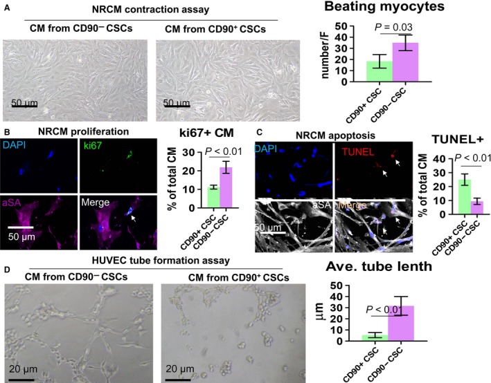
Figure 2.

In vitro cardiomyocyte and endothelial cell‐based assays. (A) NRCMs cocultured with conditioned media from CD90− or CD90+ CSCs. Scale bar = 50 μm. (B and C) Representative fluorescent micrographs showing ki67 positive (B) and TUNEL positive (C) cells in NRCM cultures. D: HUVEC tube formation on Matrigel in the presence of conditioned media from CD90− or CD90+ CSCs. Scale bar = 20 μm. Two‐tailed t‐test for comparison.
