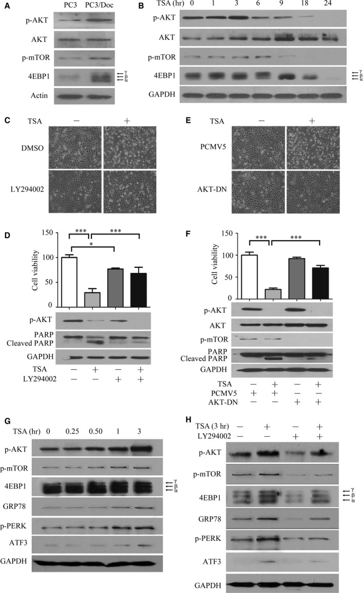
Figure 4.

TSA affects protein synthesis through modulating AKT/mTOR/4EBP1 signalling. (A) Western blot analyses of the expression differences of the AKT/mTOR/4EBP1 pathway between PC3 and PC3/Doc cells. (B) Analysis of the effect of TSA on proteins associated with protein synthesis by Western blot analysis. (C, D) The PI3K inhibitor LY294002 attenuated TSA‐mediated cell death. PC3/Doc cells were incubated with LY294002 (20 μM) for 2 hrs prior to 0.4 μM TSA treatment. Morphological changes were observed under a light microscope, cell viability was measured by the MTT assay, and cleaved caspase‐3 and PARP expression were determined by Western blot analysis. (E, F) PC3/Doc cells transfected with AKT1‐DN for 24 hrs prior to 0.4 μM TSA treatment. Cell viability was investigated by MTT (upper panel), and cell death was determined by Western blot analysis. *P < 0.05, ***P < 0.001. Data shown are the mean ± S.D., n = 3. (G) Western blot analyses of the effect of AKT/mTOR signalling and proteins associated with ER stress when exposed to TSA in 3 hrs. (H) PC3/Doc cells were incubated with LY294002 for 2 hrs prior to 3‐hrs TSA treatment. The expression of AKT/mTOR signalling and proteins associated with ER stress were determined by Western blot analysis.
