Figure 5.

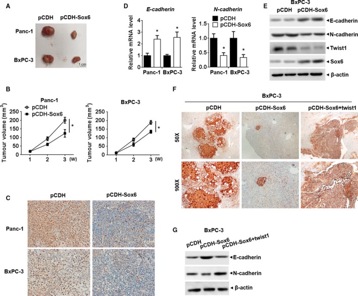
Sox6 regulated pancreatic cancer development in vivo (A). Representative images of xenograft tumours from Sox6 overexpressing or control PC cells in nude mice. (B) Size of the tumours was measured and calculated every week, completing growth curve of xenograft tumours in different groups. (C) IHC staining for PCNA in the tumour tissues developed from indicated PC cells. (D) qPCR analysis of E‐cadherin and N‐cadherin in the indicated group of tumours. (E) Western blot assay of E‐cadherin, N‐cadherin and Twist1 in the indicated group of tumours. (F) BxPC‐3 cells stably expression of Sox6 or Twist1 were produced and injected into the spleen of BALB/c nude mice under anaesthesia. Mice were killed 8 weeks after injection, and the livers were surgically excised and subjected to IHC staining for CEA to detect the liver lesion caused by PC metastasis. (G) Western blot analysis of E‐cadherin and N‐cadherin in the indicated groups of tumour. *P < 0.05.
