Figure 7.

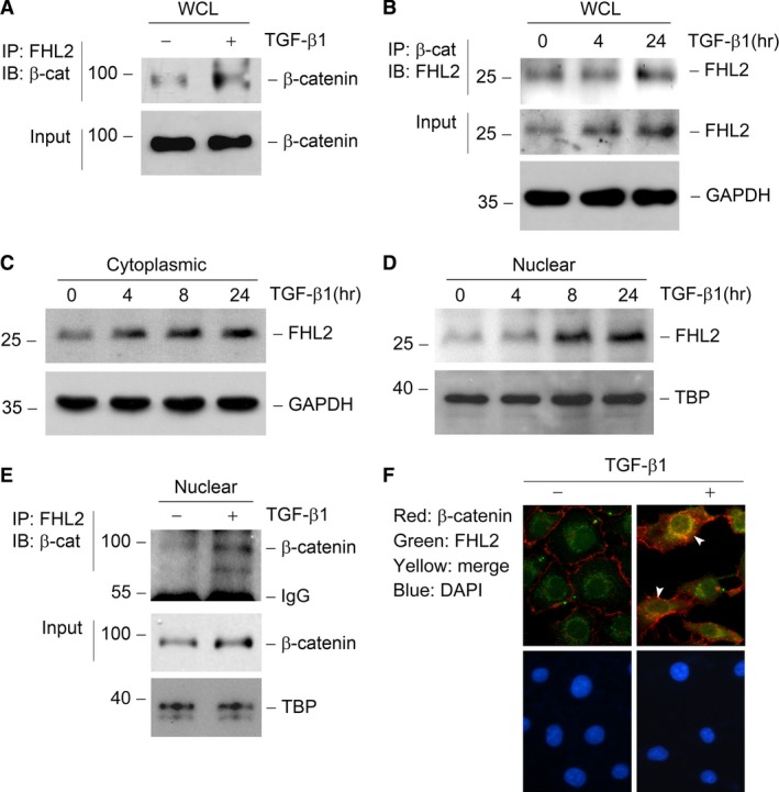
TGF‐β1 induces FHL2 to interact with β‐catenin, especially in the nuclei. (A and B) Coimmunoprecipitation (IP) reveal that TGF‐β1 induces FHL2/β‐catenin complex formation in tubular epithelial cells. Cell lysates were prepared after TGF‐β1 treatment as indicated and immunoprecipitated with specific antibody against FHL2 or β‐catenin, followed by immunoblotting for β‐catenin (A) or FHL2 (B), respectively. Cellular β‐catenin (A) or FHL2 and GAPDH (B) levels were assessed by routine Western blot analysis of whole cell lysate (WCL), respectively. (C and D) TGF‐β1 induces FHL2 expression both in the cytosol (C) and nuclei (D) in tubular epithelial cells. NRK‐52E cells were treated with 2 ng/ml TGF‐β1 for various periods as indicated, and the nuclear and cytoplasmic proteins from WCL were prepared and immunoblotted with antibodies against FHL2, TBP or GAPDH, respectively. (E) IP demonstrates a FHL2/β‐catenin complex formation induced by TGF‐β1 in the nuclei of tubular epithelial cells. The nuclear protein from WCL was prepared and immunoprecipitated with specific antibody against FHL2, followed by immunoblotting for β‐catenin. Nuclear β‐catenin and TBP levels were assessed by routine Western blot analysis of nuclear protein, respectively. (F) Immunofluorescence staining exhibits the colocalization of FHL2 and β‐catenin in tubular epithelial cells. NRK‐52E cells were treated with 2 ng/ml TGF‐β1 for 24 hrs, followed by staining with anti‐FHL2 and anti‐β‐catenin antibodies. Arrowheads indicate the colocalization sites in NRK‐52E cells. DAPI, 4′, 6‐diamidino‐2‐phenylindole‐HCl.
