Figure 1.

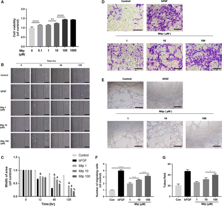
Effect of Mtp on cellular proliferation, migration, recruitment and tube formation of BM‐EPCs. (A) Cell proliferation results of BM‐EPCs treated with different concentrations of Mtp for 48 hrs. Cells proliferated evidently faster after Mtp treatment; (B, C) cell migration regulated by Mtp treatments. Scratch assay showed that EPCs migrated evidently faster in the Mtp‐treated group (scale bar: 200 μm); Data are presented as mean ± SD, & P < 0.05 versus the control group, # P < 0.05 versus the 1 μM Mtp treated group, % P < 0.05 versus the 10 μM Mtp treated group; (D, F) transwell chemotaxis assay results of BM‐EPCs with different treatments. BM‐EPCs were treated with PBS (control), 50 ng/ml bFGF and 1, 10 and 100 μM Mtp in the lower chamber for 3‐hrs incubation. Numbers of migrated cells were quantified by counting cells in 10 random fields using an inverted microscope (scale bar: 50 μm). The migration of BM‐EPCs was enhanced after Mtp treatment; (E, G) in vitro tube formation results of BM‐EPCs treated by Mtp. Cells were grown on Matrigel™ for 6 hrs under normal growth conditions, five independent fields were assessed for each well and the number of tubes were determined (scale bar: 100 μm). The tube formation ability of BM‐EPCs was improved after Mtp treatment. n = 3 independent experiments. *P < 0.05, **P < 0.01, ***P < 0.005, and ****P < 0.001 versus the indicated group.
