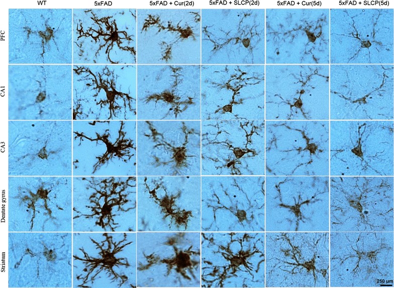
Fig. 5.

Morphological changes of single microglial cells in the 5xFAD mouse brain after treatment with Cur or SLCP. The 5xFAD mice were treated with Cur or SLCP for 2- or 5-days, sacrificed, and their brain sections were immunolabeled for Iba-1 antibody. The Iba-1-IR appeared to be more intense and the branching of microglia were more diffuse in the brain tissue of the vehicle-treated 5xFAD mice compared to those mice treated with Cur or SLCP for either 2- or 5-days. Scale bar indicates 250 µm and is applicable to all images
