Fig. 4.

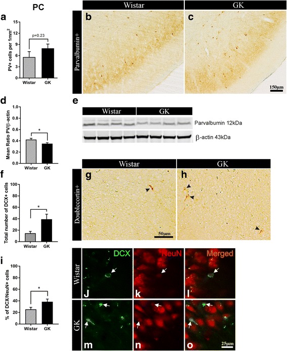
Diabetes decreases parvalbumin expression and impairs neuroblast differentiation in the piriform cortex. a Density of PV+ interneurons and, b-c representative microphotographs of PV+ staining in the PC of non-diabetic Wistar versus T2D GK rats (n = 7). d Relative level of PV normalized against beta-actin and, e representative bands showing PV expression level in the PC of non-diabetic vs. T2D rats (n = 4) (Western blot). f Total number and, g-h representative microphotographs of post-mitotic immature DCX+ neurons of embryonic origin in the PC of middle-aged Wistar vs. GK rats. i Percent of "differentiating" DCX/NeuN+ neurons and, j-o representative microphotographs of double-stained DCX/NeuN+ neurons (white arrows) in the PC of Wistar vs. GK rats (n = 7–9). Two-tailed, unpaired t-test. The data are means ±S.E.M., * p < 0.05
