Figure 1.

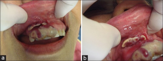
(a) Clinical image showing a tan-red exophytic, lobulated mass of the maxillary anterior facial gingiva. (b) A separate, similar appearing smaller lesion was identified in the right maxillary vestibule

(a) Clinical image showing a tan-red exophytic, lobulated mass of the maxillary anterior facial gingiva. (b) A separate, similar appearing smaller lesion was identified in the right maxillary vestibule