
Fig. 2.
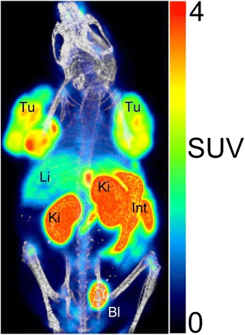
PET/CT image (three-dimensional, maximum intensity projections (MIP)) of a female CD1 nu/nu mouse. Static scan (whole body, 120–150 min p.i.) of CHL-GLP-1 tumor-bearing mouse injected with [18F]2 (12.7 MBq (1.3 nmol)). Anesthesia was maintained with 2–3% isoflurane in O2/air. SUV standardized uptake value, Tu tumor, Ki kidneys; Li liver, Bl urinary bladder, Int intestinal tract