
Fig. 4. ST3Gal6 sialylation of IAP in preventing intestinal inflammation.
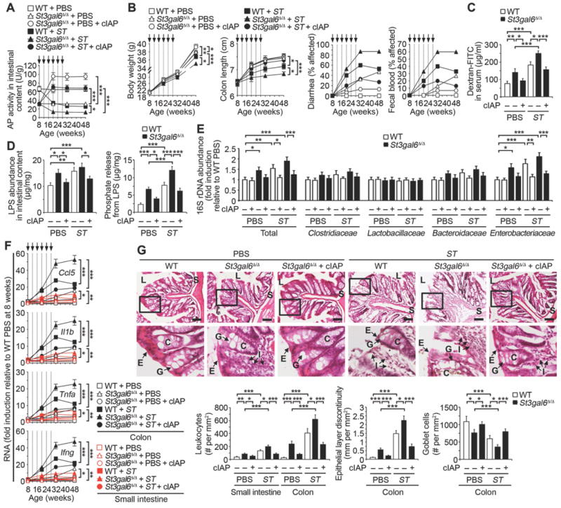
Indicated genotypes following ST re-infection (arrows) were analyzed in absence or presence of cIAP. (A) AP activity (n = 32 per condition). (B) Body weight (n = 10 per condition), colon length (n = 32 per condition), diarrhea (n = 30 per condition), and fecal blood (n = 30 per condition). (C) Intestinal epithelial barrier function. (D) LPS abundance and phosphate released from LPS of intestinal content. (E) Commensal microbiome 16S rDNA in intestinal content (n = 10 per condition). (F) Inflammatory cytokine RNA in colon and small intestine (n = 24 per condition). (G) H & E-stained colon sections. Graphs are representative of ten fields of view (four mice per condition). Scale bars: 100 µm. (C and D) Data were acquired from mice 20 weeks of age prior to fourth infection. (E–G) Data were acquired from mice 32 weeks of age and 4 weeks following the last infection. (C and D) n = 8 per condition. Error bars, means ± SEM. ***P < 0.001; **P < 0.01; *P < 0.05; one-way ANOVA with Tukey’s multiple comparisons test (A) to (G).