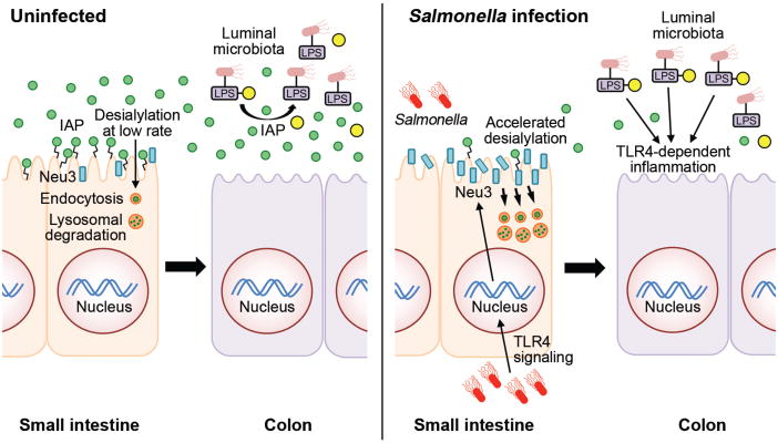
Fig. 8. Model of intestinal inflammation due to recurrent Gram-negative ST infection.

In the absence of infection of the small intestine, the anti-inflammatory GPI–linked IAP glycoprotein (green circles) is highly expressed on the enterocyte cell surface. IAP is eventually released into the lumen and travels through the intestinal tract to the colon where it detoxifies LPS-phosphate produced by Gram-negative and commensal bacteria via dephosphorylation (yellow circles). Nascent IAP at the enterocyte cell surface undergoes a low rate of desialylation linked to the rate of internalization and degradation involving a normal mechanism of IAP aging and turnover. Enterocytes of the small intestine respond to LPS-phosphate and ST infection by activating Tlr4 function, which induces host Neu3 neuraminidase (blue bars) on the enterocyte surface. Increased neuraminidase activity accelerates the rate of IAP de-sialylation and internalization (orange circles), reducing IAP abundance and resulting in increased levels of LPS-phosphate in the colon where Tlr4 activation elicits inflammation and disease.
