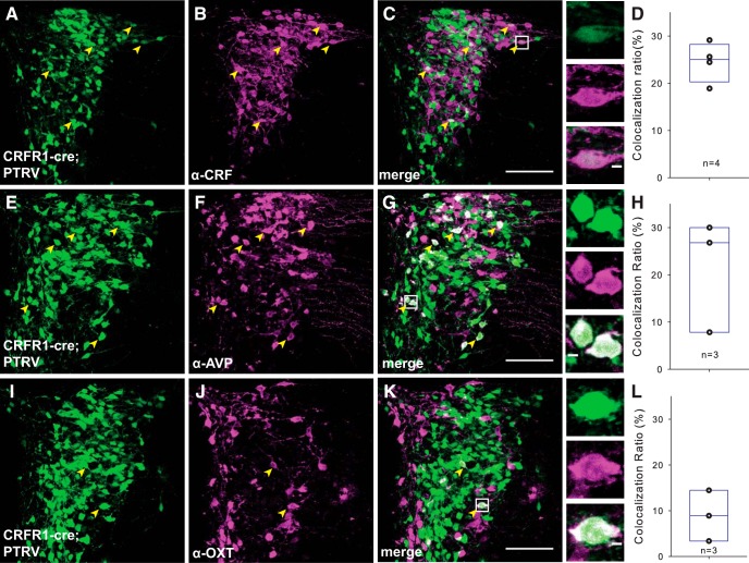
Figure 5.
PVN CRFR1 neurons receive intra-PVN inputs. A–C, In PTRV-assisted mapping of CRFR1 neuron synaptic inputs, many of the presynaptic neurons (green, left) are positive for CRF (middle panels). The merged image shows that many CRF neurons make monosynaptic connections with CRFR1 neurons. Arrowheads point to the neurons that express eGFP and are immunoreactive for CRF. Insets, Higher-power micrographs of CRF-positive PTRV traced neurons. D, Quantification of CRF+ input neurons. E–G, We also identified many presynaptic neurons (green, left) as positive for AVP (middle). The merged image shows the abundance of AVP+ magnocellular neurons that are presynaptic to CRFR1 PVN neurons. H, Quantification of AVP+ input neurons. I–K, Presynaptic OT+ neurons were also labeled by monosynaptic tracing, but were less frequent. L, Quantification of OT+ input neurons. Scale bars: 100 μm for C, G, K; 5 μm for inserts.

