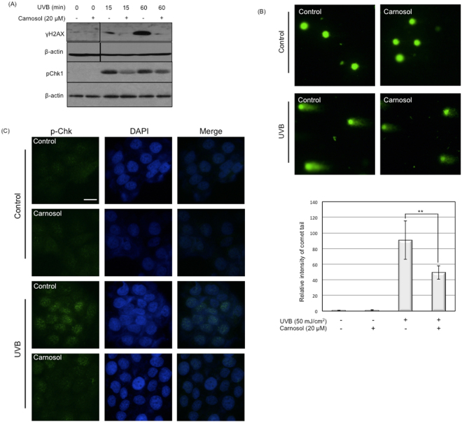
Figure 3.
Carnosol protects UVB-induced DNA damage. HaCaT cells were exposed to 50 mJ/cm2 UVB radiation in the presence or absence of carnosol (20 μM). (A) Cells were lysed at indicated time point and protein levels of phosphorylated H2AX (γH2AX) and Chk1 (p-chk1) were determined by western blot. The data represents three sets of independent experiments. (B) Comet assay for HaCaT exposed to UVB radiation. After cell treatment, cells were trypsinized and collected for comet assay. The tail intensity was semi-quantitatively analyzed using Image J. *p < 0.05, **p < 0.01. (C) Cells were immunostained with p-Chk1 upon UVB radiation with or without carnosol (20 μM) treatment. p-Chk1 was stained with green fluorescent dye, while nucleus was stained with DAPI. Scale indicates 20 μm.

