Figure 5.
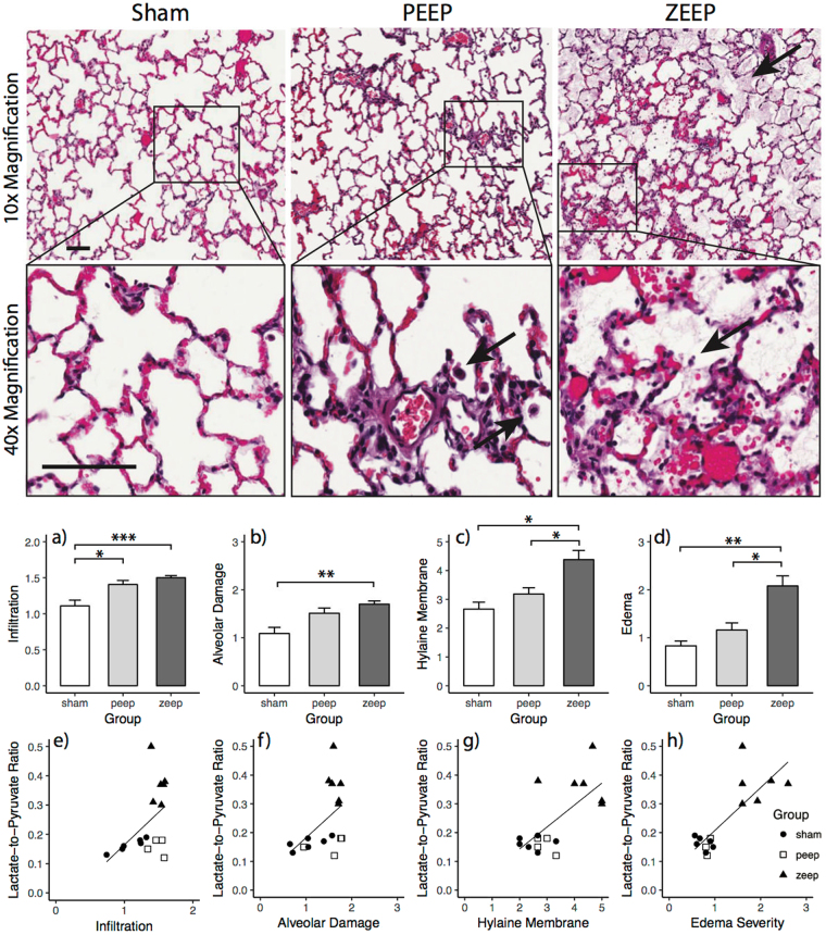
(Top) Hematoxylin and Eosin (H&E) stained lung sections of rats from each group. Representative images shown are at 10× and 40×. In ZEEP lungs, arrows show the presence of fluid within the alveolar space. Solid arrows delineate infiltrates and debris. In lungs subjected to PEEP, alveolar structure is more intact. Infiltrates are shown by solid arrows. The scale bar is 100 µm. (Middle) Average injury scores for various morphological features for different groups. ANOVA showed a significant difference among groups (infiltration: χ2(2) = 11.273, p = 0.003, alveolar damage: χ2(2) = 10.000, p = 0.007, hyaline membrane deposition: χ2(2) = 10.678, p = 0.005 and edema: χ2(2) = 12.154, p = 0.002). Infiltration (a) and alveolar damage scores (b) were significantly higher in the ZEEP group than in the sham group (p = 0.001, p = 0.012), while infiltration was significantly higher in the ZEEP group than the PEEP group (p = 0.035). Hyaline membrane deposition (c) and edema severity (e) were both significantly higher in the ZEEP group relative to the PEEP (p = 0.048, p = 0.041) and sham (p = 0.014, p = 0.006) groups. (Bottom) Among the histological features, hyaline membrane deposition (r = 0.612, p = 0.003) (g) and edema severity (r = 0.663, p = 0.001) (h) were strongly correlated with lactate-to-pyruvate ratio. (*p < 0.05, **p < 0.01, ***p < 0.001).

