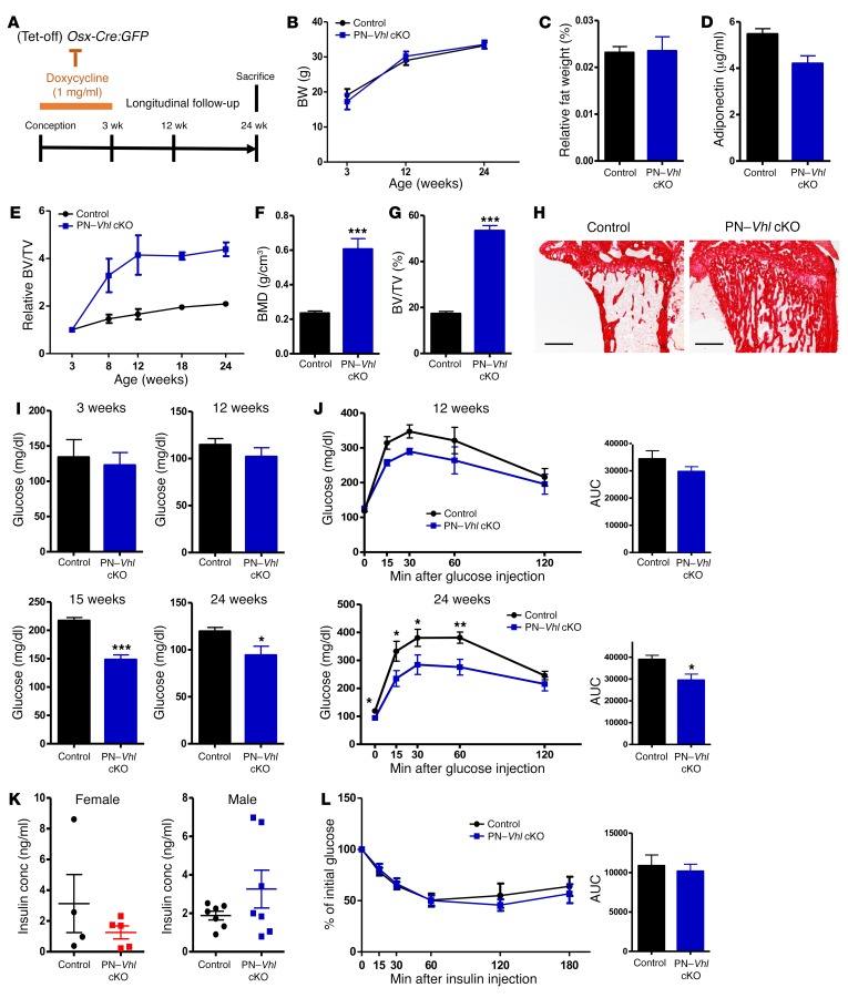
Figure 4. PN–Vhl cKO mice recapitulate the key skeletal and systemic features of the constitutive Vhl cKO model while showing normal BW and fat mass.
(A) Scheme of doxycyclin administration to silence Osx-Cre:GFP activity from conception until weaning. (B) BW of control and PN–Vhl cKO mice at the indicated ages (n = 6–9). (C) Abdominal fat weight as percentage of BW at 24 weeks (n = 6–7). (D) Serum adiponectin levels at 24 weeks (n = 7–8). (E) Relative BV/TV change over time analyzed by in vivo micro-CT. (F and G) Bone mineral density (BMD) (F) and BV/TV (%) (G) determined by ex vivo micro-CT at 24 weeks (n = 7–8). (H) Representative sirius red–stained tibia sections (scale bars: 500 μm) (n = 3). (I) Blood glucose levels at 3 weeks of age (overnight fast, n = 6), 12 weeks (overnight fast, n = 6–8), 15 weeks (3-hour fast, n = 6–8), and 24 weeks of age (overnight fast, n = 7–8). (J) GTT at 12 weeks (n = 3–7) and 24 weeks (n = 7–8), with corresponding AUC calculations. (K) Serum insulin levels in random-fed conditions at 24 weeks in females (left, n = 4–5) and males (right, n = 7). (L) ITT and corresponding AUC (n = 4–5). Graphs represent mean ± SEM, and *P < 0.05, **P < 0.01, ***P < 0.001 by Student’s t test between genotypes, unless indicated otherwise. All data in B–J and L were obtained in male control and induced PN–Vhl cKO mice. Corresponding data from female groups are shown in Supplemental Figure 8.

