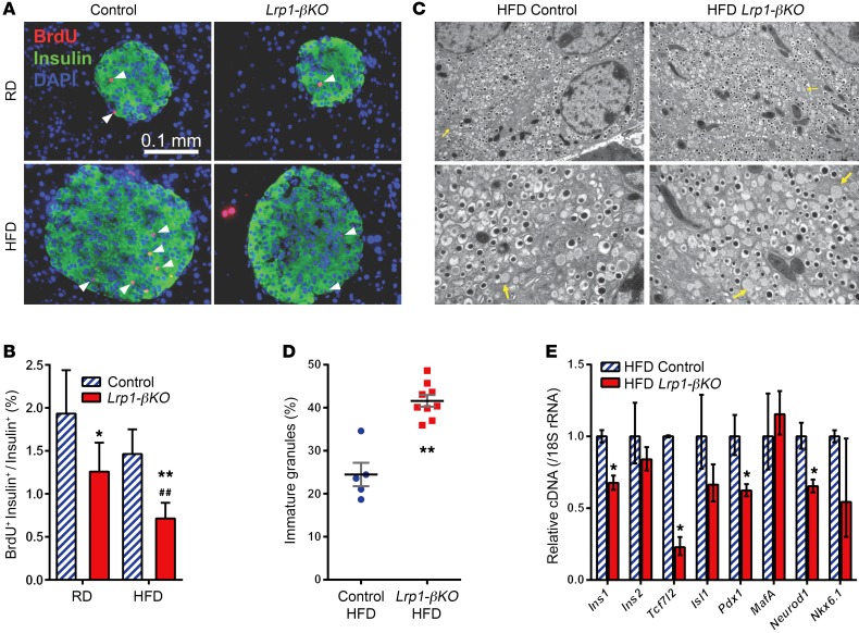
Figure 3. Defective proliferation and insulin production in LRP1-KO β cells after HFD.
(A and B) Four months after doxycycline treatment, mice were subjected to 3 consecutive daily i.p. injections of BrdU, and their pancreas sections were subjected to immunofluorescence of BrdU (red), merged with insulin (green) and DAPI (blue). (A) Representative images. Arrowheads show examples of BrdU-positive β cells. (B) Percentage of BrdU-positive β cells. n = 2.8–8.1 × 10³ cells (31–48 islets) per condition. Data are presented as percentage ± 95% CI. *P < 0.05; **P < 0.01 for Lrp1-βKO versus control mice; ##P < 0.01 for RD versus HFD by Z test. (C and D) Transmission electron microscopy of β cells in mice after 8 months of HFD. (C) Representative fields. Original magnification, ×10,000 (upper panels); ×25,000 (lower panels). Arrows show examples of immature secretory granules. (D) Percentage of granules lacking an electron-dense core. n = 5 (control); n = 9 (Lrp1-βKO) ×25,000 fields, with 75–220 granules per field. (E) RT-qPCR of insulin and β cell transcription factors with pancreatic islets from mice after 8 months of HFD. n = 3 to 4 mice per genotype. Data are presented as mean ± SEM. *P < 0.05; **P < 0.01 for Lrp1-βKO versus control mice by 2-tailed unpaired Student’s t test.

