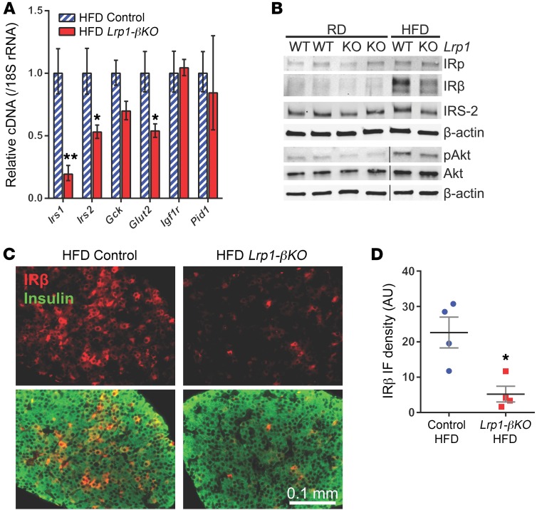
Figure 4. Diminished insulin signaling in Lrp1-βKO islets after HFD.
Experiments used mice 8 months after doxycycline treatment. (A) RT-qPCR of insulin/IGF signaling genes with pancreatic islets. n = 3 to 4 mice per genotype. (B) Representative Western blots of insulin-signaling molecules with pancreatic islets. Noncontiguous lanes run on the same gel are separated by black lines. (C and D) Immunofluorescence of IRβ on pancreas sections. (C) Representative images. Upper panels: IRβ signal (red) only. Lower panels: merged with insulin (green). (D) IRβ signal intensity in insulin-positive cells is quantitated in individual mice (4–6 ×20 sections per mouse). Data are presented as mean ± SEM. *P < 0.05; **P < 0.01 for Lrp1-βKO versus control mice by 2-tailed unpaired Student’s t test.

