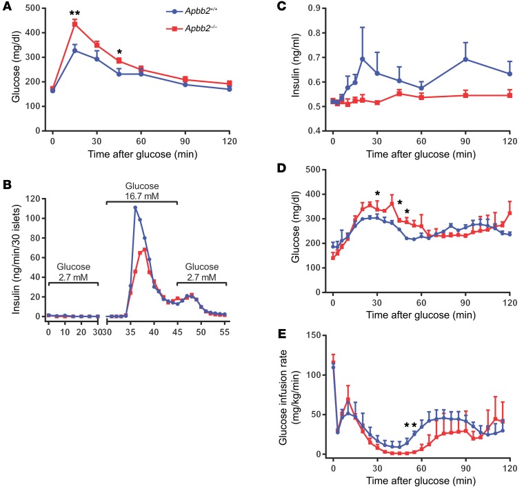
Figure 9. Apbb2 deletion impairs β cell function in RD-fed mice.
Adult Apbb2-KO mice (Apbb2–/–) and WT littermates (Apbb2+/+) were fed on RD. (A) Blood glucose during i.p. glucose tolerance test (1 mg/g body weight). n = 6 (Apbb2+/+) and n = 9 (Apbb2–/–) mice. (B) Thirty islets per genotype were subjected to perifusion with glucose concentration at 2.7 mM (0–30 minutes), 16.7 mM (30–45 minutes), and 2.7 mM (45–55 minutes). Insulin concentrations of perifusion fractions were assayed and presented. (C–E) Plasma insulin (C), blood glucose (D), and glucose infusion rate (E) during hyperglycemic clamp. n = 3 mice per genotype. Data are presented as mean ± SEM. *P < 0.05;**P < 0.01 for Apbb2–/– versus Apbb2+/+ mice by 2-tailed unpaired Student’s t test.

