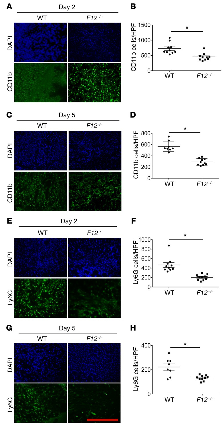
Figure 1. FXII influences leukocyte migration into skin wounds.
(A and C) Frozen sections of day 2 (D2) and day 5 (D5) wounds were stained with anti-CD11b antibody to assess leukocyte infiltration. (B and D) Total number of CD11b cells per high-power field (HPF). WT, n = 10; F12–/–, n = 10 mice (B); WT, n = 8; F12–/–, n = 10 mice (D). (E and G) Representative frozen sections from day 2 and day 5 wounds were stained with anti-Ly6G antibody to determine neutrophil infiltration. (F and H) The numbers of Ly6G cells/high-power field are shown. WT, n = 10; F12–/–, n = 10 mice (F); WT, n = 8; F12–/–, n = 10 mice (H). Fluorescent images were obtained using a Nikon TE2000-S microscope at ×20 magnification. CD11b and Ly6G staining in all panels was compared by morphometric analysis of cell number per high-power field using ImageJ software (NIH). Data represent mean ± SEM. *P < 0.01 vs. WT control mice by Student’s t test. Scale bar: 50 μm.

