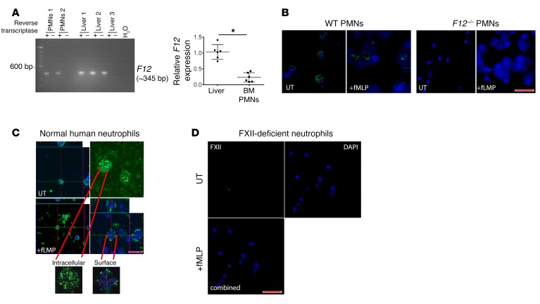
Figure 5. Neutrophils (PMNs) express F12 mRNA and FXII.
(A) Left panel: murine total mRNA was isolated from BM-derived neutrophils (PMNs 1,2) or homogenized livers (Liver 1–3), and first-strand cDNA was synthesized with SuperScript III reverse transcriptase. The PCR product from the same animal on an exon 1–6 probe is shown. Images are representative of 3 experiments. Right panel: relative F12 expression in liver and BM-derived neutrophils as determined by 2–ΔΔCt. Mean ± SEM. *P = 0.0001, Student’s t test. (B) Isolated WT (left panel) and F12–/– (right panel) murine peripheral blood neutrophils were incubated with media or fMLP for 2 hours. Cells were stained with primary antibody directed against FXII (green); nuclei were counterstained with DAPI (blue). Images are representative of n = 3 experiments. Original magnification, ×20. Scale bar: 10 μm. (C) Confocal visualization of FXII in normal human peripheral neutrophils. Neutrophils were incubated with media (UT; top panels) or fMLP (bottom panels), then either not permeabilized (surface) or permeabilized (intracellular) and stained with antibody to FXII. Confocal microscopy indicates the intracellular location of FXII protein before treatment and trafficking to the cell surface after stimulation with fMLP. Images shown are representative of 3 individual experiments. Original magnification, ×20 (left panel); ×100 (right panel). Scale bar: 5 μm. (D) Confocal FXII visualization in neutrophils from FXII-deficient patient in UT (top) and fMLP-treated (bottom) cells. FXII is absent in peripheral neutrophils from a patient with congenital FXII deficiency. Original magnification, ×10. Scale bar: 10 μm.

