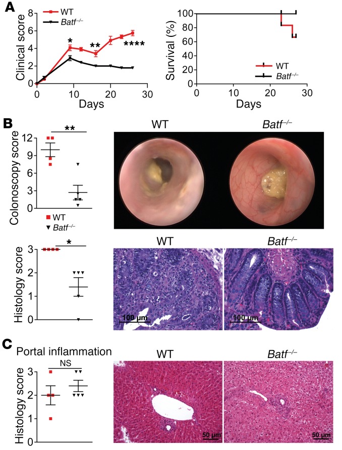
Figure 4. BATF-expressing T cells control intestinal but not hepatic GVHD in a miHA-mismatched model.
(A) Clinical GVHD score and survival rates following transfer of allogeneic WT (red squares) and Batf –/– (black triangles) CD3+ C57Bl/6 donor T cells into irradiated H-2b+ BALB.b mice after transplantation of T cell–depleted CD45.1+ B6.SJL WT BM. Results from 1 representative experiment are shown (n = 6 WT and n = 5 Batf–/– mice). (B) Representative endoscopic (upper row) and histologic cross-sectional (lower row) images showing GVHD-associated colitis activity during established GVHD on day 27. Scatter plots summarize the pooled results of colonoscopy and histology scores for WT (n = 4) and Batf–/– (n = 5) T cell–recipient mice. Scale bars: 100 μm. (C) Histopathologic evaluation of GVHD-associated liver lesions (scatter plot) assesses portal inflammation of the liver, and representative images of histopathologic cross-sections are shown. Scale bars: 50 μm. Data represent the mean ± SEM. *P < 0.05, **P < 0.01, and ****P < 0.0001, by 2-sided, unpaired Student’s t test.

