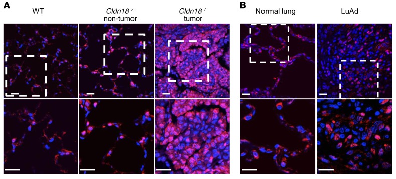
Figure 7. Nuclear YAP expression in lung adenocarcinoma (LuAd).
(A) Nuclear YAP (red) is present in Cldn18–/– mouse lungs in areas both with and without tumor. Lower panel shows higher magnification views of rectangle in upper panel. DAPI is the nuclear counterstain. n = 2. Scale bars: 20 μm. (B) Nuclear YAP is increased in human LuAd compared with normal lung. Lower panel shows higher magnification views of rectangle in upper panel. DAPI is the nuclear counterstain. n = 2. Scale bars: 20 μm.

