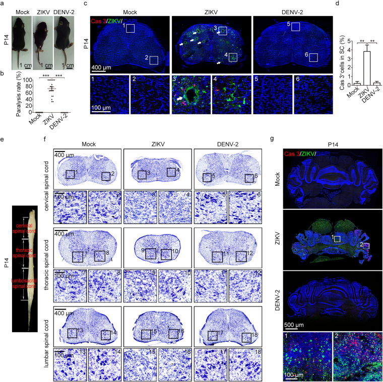
Figure 5.
Severe movement defects were induced by maternal ZIKV infection. (a) Representative images of P14 offspring mice. The hind limbs of ZIKV-infected mice were paralyzed. (b) Quantification of the paralysis rate during P0-P28. (c) Representative images of lumbar spinal cords stained for Cas3 and ZIKV. White arrows, cavities. (d) Percentage of Cas3-positive cells within lumbar spinal cords. nMock = 4, nZIKV = 8, nDENV-2 = 4 samples from three independent experiments. (e) Representative image of a P14 spinal cord to illustrate the different segments. (f) Nissl staining of P14 cervical, thoracic, and lumbar spinal cord. (g) Representative images of the cerebellum stained for Cas3 and ZIKV. Quantification data are presented as the mean ± SEM. One-way ANOVA with Bonferroni’s post hoc test, **p < 0.01, ***p < 0.001.

