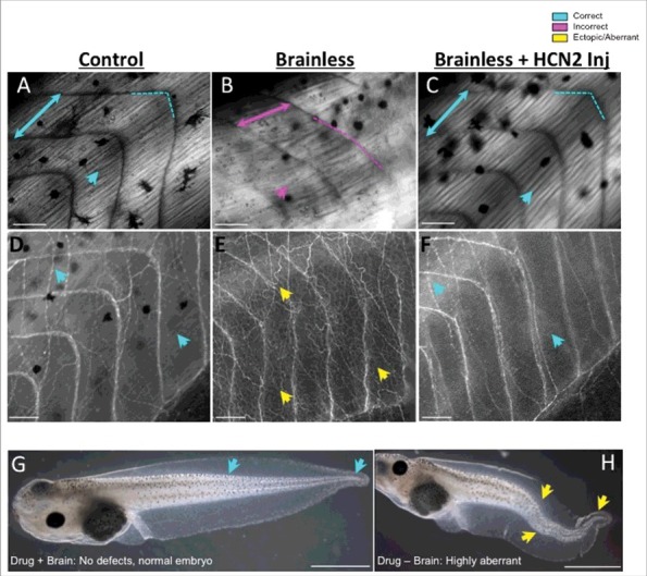
Figure 2.

Functions of the very early brain include guiding and protecting correct embryo morphogenesis, which can be mimicked by ectopic expression of ion channels. Comparative images of muscle (A-C, under polarized light) and nerve (D-F, revealed by immunofluorescence against acetylated-alpha tubulin) structures of embryos that developed with a brain (Control), embryos that developed without a brain (Brainless), and embryos that developed without a brain and were injected with messenger RNA encoding the HCN2 ion channel (Brainless + HCN2 Inj). The absence of a brain during development leads to abnormal development of the muscles and the peripheral nerves, at considerable distances from the head, with disorganization of the body plan, myotomes lacking proper angles (magenta-dashed line in B), alterations in somite length (represented by two-headed arrows) and ectopic growth of nerve tissue (yellow arrows in E). By manipulating bioelectricity, for example via the mis-expression of HCN2, rescues the devastating muscle and nerve mispatterning that occurs in brainless animals (turquoise arrows in C, F). Rostral is right and dorsal is up. Scale bar = 100 μm. G, H. The effects of the drug (RS)-(tetrazol-5-yl)glycine during development depends on the presence or absence of the nascent brain in the embryo. While exposure to this drug does not cause developmental defects in control embryos (turquoise arrows in G indicate correct tail phenotype), it leads to severe deformities (yellow arrows in H demarcate bent spinal cord and tail aberrations) when the brain is not present to protect. Rostral is left and dorsal is up. Scale bar = 1 mm.
