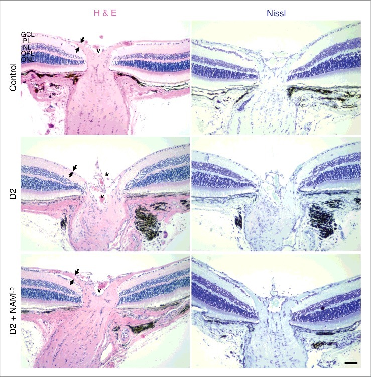
Figure 2.

NAM prevents optic nerve cupping in glaucoma. The presence of optic nerve cupping was assessed using haematoxylin and eosin staining (H & E) and cresyl violet staining (Nissl). In control eyes (D2-Gpnmb+) there is a robust ganglion cell layer and nerve fiber layer (which contains axons from the retinal ganglion cells, denoted by the distance between the black arrows). In D2 eyes that have undergone glaucomatous neurodegeneration there is evident optic nerve cupping (asterisk), and a loss of ganglion cell layer cells and nerve fiber layer thickness. Cupping and RGC loss were absent in eyes of NAM treated mice (D2 + NAMLo), thus NAM treatment protected from all assessed signs of glaucoma. Eyes were assessed at 12 months of age. V corresponds to the central retinal vessel. GCL = ganglion cell layer, IPL = inner plexiform layer, INL = inner nuclear layer, OPL = outer plexiform layer, ONL = outer nuclear layer. Scale bar = 50 μm.
