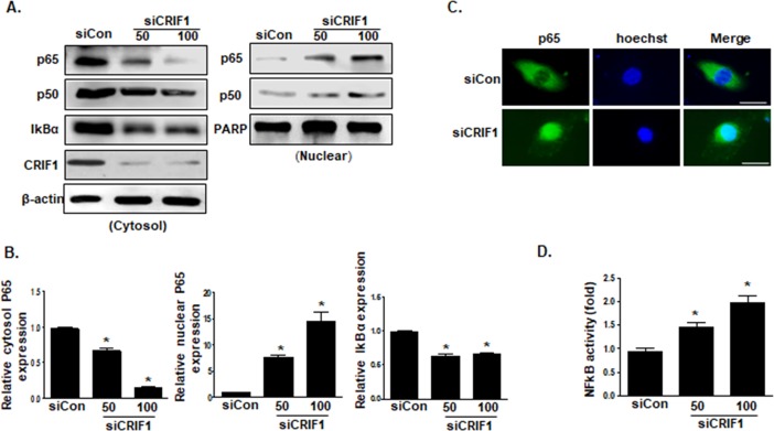
Fig 3. CRIF1 deletion stimulated the activation of the NF-κB signaling pathway in HUVECs.
(A) HUVECs were transfected with CRIF1 (50 or 100 pmol) for 48 h, cytosolic and nuclear fractions were isolated, and p65 and IκBα protein levels in the cytoplasm and nucleus were detected by western blotting. (B) p65 and IκBα protein levels were quantified by densitometric analysis. (C) Representative immunofluorescence images of p65 (green) and Hoechst (blue) staining. Scale bars indicate 20 um. (D) NF-κB activity was measured using a dual luciferase reporter gene assay in HUVECs transfected with CRIF1 siRNA (50 and 100 pmol) for 48 h. The graph presents the fold changes in NF-κB activation compared with that in the control. All western blots are representative of three independent experiments. The data are presented as means ± SEM of three independent experiments. *p < 0.05 vs. control cells.

