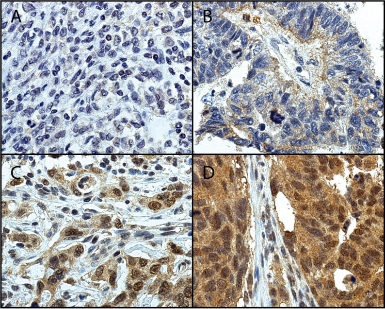
Fig 1. Representative images of UCHL5 staining in gastric cancer.

Tumors with A) negative (0), B) weak positive (1), C) moderate positive (2), and D) strong positive (3) staining. Original magnification 40x.

Tumors with A) negative (0), B) weak positive (1), C) moderate positive (2), and D) strong positive (3) staining. Original magnification 40x.