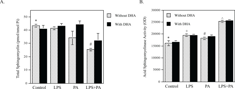
Fig 3. DHA has no effect on SM hydrolysis and ASMase activity stimulated by LPS and PA.
A. RAW264.7 macrophages were treated with 1 ng/ml of LPS, 100 μM of PA or both 1 ng/ml LPS and 100 μM of PA in the absence or presence of 100 μM of DHA for 12 h. After treatment, cellular sphingomyelin was quantified using lipidomics. * vs. #, p<0.01. B. RAW264.7 macrophages were treated with 1 ng/ml of LPS, 100 μM of PA or both 1 ng/ml LPS and 100 μM of PA in the absence or presence of 100 μM of DHA for 2 h. After treatment, cellular ASMase activity was determined as described in the Methods. * vs. +, p<0.05; * vs. #, p<0.05; * vs. ^, p<0.05.

