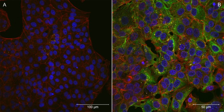
Fig 3.
Laser scanning confocal microscopy of Rickettsia helvetica AS819-infected Vero E6 cells five days (A) and 20 days (B) after infection. F-actin was stained with Alexa Fluor 568 Phalloidin (red), nuclei were stained with DAPI (blue), and rickettsiae are coloured yellow (magnification 40x). The bar indicates 100 μm (a) and 50 μm (b).

