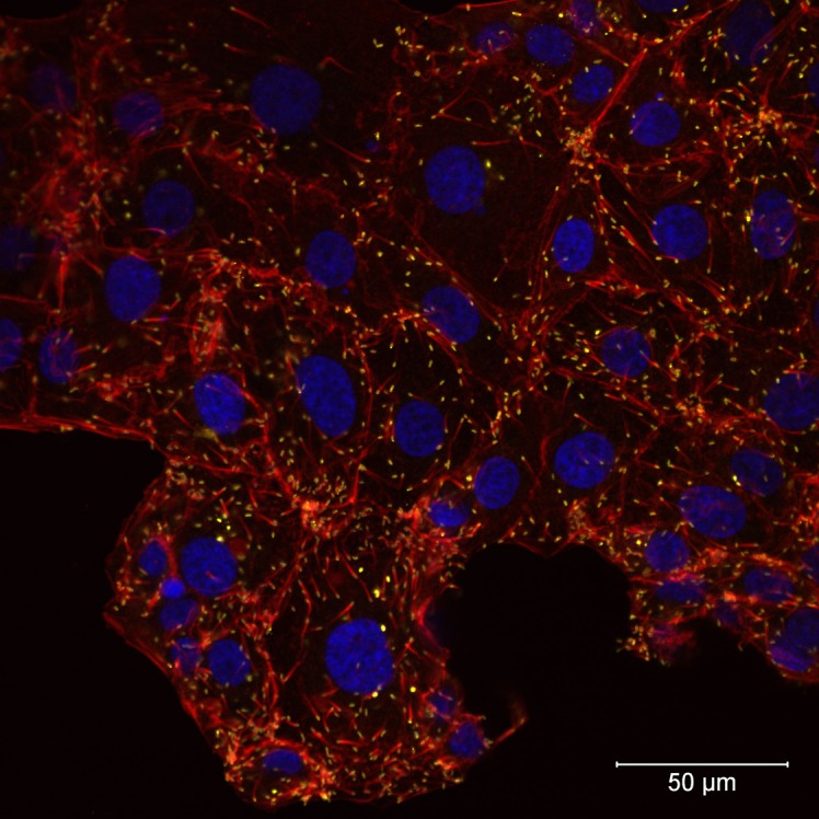
Fig 5. Laser scanning confocal microscopy of Rickettsia conorii VR141-infected Vero E6 cells four days after infection.

Actin tails at one pole of R. conorii VR141 can be seen. F-actin was stained with Alexa Fluor 568 Phalloidin (red), nuclei were stained with DAPI (blue), and R. conorii VR141 is presented in yellow (magnification 40x). The bar indicates 50 μm.
