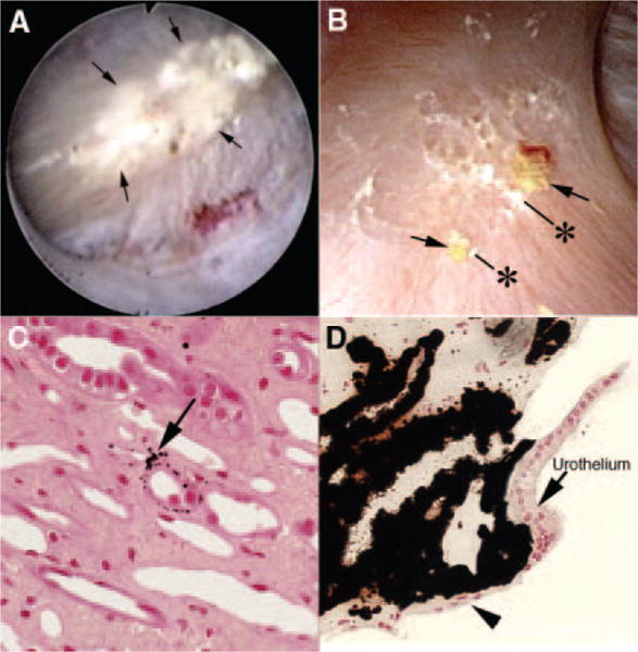
Figure 1. Endoscopic and histologic characteristics of interstitial plaque in idiopathic calcium oxalate stone-formers (ICSFs).

(A) Example of a papilla from an ICSF that was video recorded at the time of the mapping protocol. The papillary tip is covered by multiple sites of an irregular white material (arrows) termed “interstitial” or “Randall’s plaque” that is seen through the urothelial covering. These regions of plaque are sites for stone attachment. (B) Two small stones (asterisks) of approximately 0.5 mm are seen associated with a region of interstitial plaque (arrows). Histologic examination of papillary biopsies taken adjacent to sites of interstitial plaque shows that crystalline material (arrow in C) initially accumulates in the basement membranes of the thin loops of Henle. With time, these small sites of deposits appear to accumulate in the interstitial space as dense islands of mineral proceeding to the basal surface of the urothelial covering of a renal papilla (arrowhead in D). Adapted from references 2,8.
